3D printing technology is considered a revolution in manufacturing. Based on the development of computer technology, the progress of production technology and the advent of new materials, 3D printing has made great progress in many fields including life sciences in the field of biomedicine. The development process is from the early exploratory preoperative model replication to the successful printing of conformal prosthesis to repair bone defects, and now a variety of new medical materials and even cell preparation technology, has made a gratifying application in the field of medical restoration. progress. The Antarctic Bear will introduce the research and development of 3D printing technology in the field of biomedicine in recent years, and introduce its research and development status in the field of Otolaryngology Head and Neck Surgery.
3D printing technology is a revolutionary, digital manufacturing technology based on pre-designed, layer-by-layer addition of specific materials. The technology was first proposed by the Massachusetts Institute of Riding in the 1980s, just like many groundbreaking technologies. Early 3D printing technology was not promoted due to complex processes, inefficiencies, expensive equipment, and rough finished products. In recent years, with the advancement of computer technology, with the development of new design software and the application of new materials and new processes, 3D printing technology has been rapidly developed in many fields.
The basic principles, manufacturing processes and main types of 3D printing technology
According to the principle of "segment manufacturing, layer by layer superposition". The 3D printing workflow is divided into 3 steps.
Step 1 is for image acquisition. The image data of the human body is obtained by optical scanning or holography, and the in vivo part can obtain high-resolution and high-contrast medical image data by CT, MRI or even positron emission tomography (PET).
The second step is image post processing. Optical scanning or holography can be obtained in a standard 3D model file (stero lithography, STL) format; data acquired by medical imaging such as CT, MRI, PET, etc. are usually stored as digital imaging and communications in medicine (DICOM). Format, this format file is read after data conversion, to achieve 3D segmentation and visualization of the target object, and finally output to the 3D printing device by computer aided design (CAD) model.
The third step is rapid prototyping. The CAD data is transformed into a three-dimensional actual object, and finally computer aided manufacturing (CAM) is realized.
.jpg)
According to the principle of molding technology and the printing materials, 3D printing technology can be subdivided into several modes: stereolithography apparatus (SLA), fused deposition modeling (FDM), selective laser Selective laser sintering (SLS), laminated object manufacturing (LOM), inkjet printing, and the like. In practical applications, different printing modes will be selected depending on the difference in printing materials and the difference in printing accuracy and prosthetic strength.
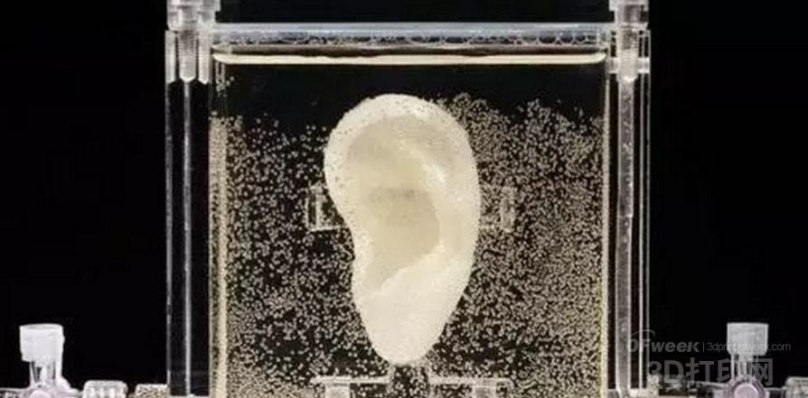
Research and application of 3D printing in the field of otolaryngology head and neck surgery
1. Ear surgery anatomical training and medical education: The humerus is the most complicated skeletal system in the human body. It not only has intricate bone and soft tissue structures, but also includes many fine gas and liquid gaps. It includes important structures such as facial nerve, internal jugular vein, cochlea, vestibule and semicircular canal. Due to the large individual differences in the structure of the tibia, anatomical variations occur. Proficiency in sacral anatomy is an indispensable skill for otologists. However, due to its irregular three-dimensional shape and fine and complex microstructure, traditional two-dimensional image teaching has considerable limitations, and the anatomical material (corpse head) is extremely scarce. The training of the physical humerus anatomy is difficult to promote. For a long time, mastering the anatomy of sacral surgery has always been a bottleneck restricting the development of otologists' skills.
Since the advent of 3D printing technology, the field of surgical anatomy training has made great progress. Vorwerk and Begall used the sacral CT scan data, combined with CAD technology, to create the earliest human humerus model using SLA technology and used for basic microdissection exercises and surgical manipulation simulations. Subsequent Suzuki et al. used SLS technology to create a more detailed tibia model through high-resolution CT scans, and anatomical and surgical procedures under the microscope using conventional surgical instruments. In 2012, Monfared et al. of the University of Washington reported that a high-fidelity and low-cost surgical middle ear model was produced by 3D printing technology, and different materials were used to simulate bone and soft tissue. The model was refined, authentic and mechanically responsive. The area performed well and was recognized in the industry and recommended to be included in the residency training guide. In addition, 3D printing can also accurately magnify the microstructure. German Hamburg Wulf and other high-resolution μCT acquisition of human hearing bones, using this technology to make a 20-fold magnified plastic anatomical model, to achieve a perfect and accurate display of the teaching structure of the small bone structure.
2, pre-operative virtual reality and 3D modeling : 3D printing technology can also be used for surgical juice or virtual modeling, that is, "painting rehearsal" before surgery. Surgeons will use the same size organs and parts that they are about to perform surgery to perform operational training, so that the surgeon has "thickness in the chest" in advance. 3D surgery exercises can shorten the operation time, find out the problems that may occur during the operation and predict the surgical outcome, avoid potential risks, and indeed improve the quality and safety of the operation. For example, the Otolaryngologist at the University of Oulu in Finland extracted the high-resolution CT data of the tibia before the complicated cochlear implant. The SLA technique was used to replicate the tibia model, which accurately showed the structures of the facial nerve canal, elliptical window and round window. Preoperative simulations were performed to reduce the risk of surgery. In addition, surgical modeling helps doctors make the necessary preoperative communication to patients and their families.
.jpg)
3. Filling and repairing of bone defect: The repair of the large hard tissue of the maxillofacial region (such as the jaw and maxilla) is usually performed with the autogenous humerus or humerus, and the drawback of "removing the east wall to make up the west wall" is not It goes without saying. With the deepening of artificial bone research, 3D printing technology has shown unique advantages in dealing with complex curved surfaces and different shapes of human bones. In addition to directly printing high biocompatible titanium callus and adjusting the particle size of the shelf material according to the porosity, the porosity and micro-aperture can be adjusted by changing the filling method of the slice to construct an adaptive cell. Active scaffolds that grow. The mandible, vertebrae, etc. have been successfully produced and used initially in clinical practice.
Using computer-aided design, the individualized titanium-titanium complex with the same normal shape as the patient's mandibular defect area was designed and implanted. The facial shape of the patient returned to normal after operation, and the occlusal relationship and mouth opening degree recovered well. The BIOMED Institute of Hasselt University in Belgium uses SLS technology to accurately fuse the layer-by-layer superimposed titanium powder particles with a numerically controlled laser to print the mandible with the same shape, supplemented by a surface-sprayed bioceramic layer to reduce the rejection reaction; A Dutch surgeon has successfully implanted a 3D printed titanium mandible for an 83-year-old patient. Since hydroxyapatite (HA) has excellent osteoinductive properties, HA is used together with a photosensitive polymer as a raw material for preparing a biologically active bone tissue engineering scaffold. Matsuo et al. of Tokyo Medical University in Japan used poly-L-lactic acid/HA (PLLA/HA) as raw material to prepare absorbable porous brackets using SLA technology, and assisted dental implant materials together for mandibular resection of mandibular tumors. Reconstructed and achieved better repair than metal titanium stents.
Home accessories are furniture items which are easy to replace and easy to move, and include almost any items that are not strictly functionally necessary in a decorated space. These accessories include such items as curtains, sofa sets, cushions, tablecloths and decorative craft products, decorative wrought iron, and so on. These items are commonly used in indoor furnishings and layout and can include cloth items, paintings, and plants.

The first thing that matters when it comes to sofas with back support is the type of the sofa. Namely, there are plenty of different types of sofas on the market, but, not all of them are for back support. Anyway, here are some of the best sofas for back support.

Home accessories, as movable decorations, reflect the owner's taste and create a personal atmosphere where they are placed. These items can break the boundaries of the traditional decoration industry, using handicrafts, textiles, collectibles, and things such as lamps, floral items, and plants re-combined to form a new concept. Home accessories vary according to the size and shape of the room space, the owner's living habits, hobbies, tastes, and their financial situation.
Home Decor,Flower Pot Rack,Room Divider Furniture,Customized Photo Frames
Jinan Tri-Tiger Technology Development Co., Ltd , https://www.tigerwoodproduct.com